Search Thermo Fisher Scientific
Disclaimer

Clicking the images or links will redirect you to a website hosted by BenchSci that provides third-party scientific content. Neither the content nor the BenchSci technology and processes for selection have been evaluated by us; we are providing them as-is and without warranty of any kind, including for use or application of the Thermo Fisher Scientific products presented.
")
 in WB")
Product Details
PA1-41658
Product Specifications
Species Reactivity
Human,
Mouse,
Rat
Host/Isotype
Rabbit
/ IgG
Class
Polyclonal
Type
Antibody
Immunogen
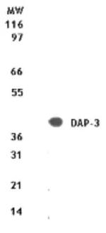
Synthetic peptide corresponding to residues 44-57 (T(44) S D S D P A K H G E Q H E(57) of mouse DAP-3.
Conjugate
Unconjugated
Form
Liquid
Concentration
0.5 mg/mL
Purification
Protein G
Storage buffer
PBS
Contains
0.02% sodium azide
Storage conditions
Store at 4°C short term. For long term storage, store at -20°C, avoiding freeze/thaw cycles.
Shipping conditions
Wet ice
RRID
AB_2090240
Product Specific Information
Suggested positive control: Jurkat, antigen standard for DAP3 (transient overexpression lysate), Jurkat whole cell lysate.
Target Information
Mammalian mitochondrial ribosomal proteins are encoded by nuclear genes and help in protein synthesis within the mitochondrion. Mitochondrial ribosomes (mitoribosomes) consist of a small 28S subunit and a large 39S subunit. They have an estimated 75% protein to rRNA composition compared to prokaryotic ribosomes, where this ratio is reversed. Another difference between mammalian mitoribosomes and prokaryotic ribosomes is that the latter contain a 5S rRNA. Among different species, the proteins comprising the mitoribosome differ greatly in sequence, and sometimes in biochemical properties, which prevents easy recognition by sequence homology. This gene encodes a 28S subunit protein that also participates in apoptotic pathways which are initiated by tumor necrosis factor-alpha, Fas ligand, and gamma interferon. This protein potentially binds ATP/GTP and might be a functional partner of the mitoribosomal protein S27. Splice variants that differ in the 5' UTR have been found for this gene; both variants encode the same protein. Pseudogenes corresponding to this gene are found on chromosomes 1q and 2q.
For Research Use Only. Not for use in diagnostic procedures. Not for resale without express authorization.
References (0)
Have you cited this product in a publication?
Let us know so we can reference it here.
Bioinformatics
Protein Aliases: 28S ribosomal protein S29, mitochondrial; DAP-3; Death-associated protein 3; DKFZp686G12159; Ionizing radiation resistance conferring protein; MGC126058; MGC126059; mitochondrial 28S ribosomal protein S29; Mitochondrial small ribosomal subunit protein mS29; MRP-S29; S29mt
Gene Aliases: 4921514D13Rik; bMRP-10; DAP-3; DAP3; MRP-S29; MRPS29; S29mt
UniProt ID: (Human) P51398, (Mouse) Q9D5V9
Entrez Gene ID: (Human) 7818, (Rat) 295238, (Mouse) 65111
Molecular Function:
![]() ribosomal protein
ribosomal protein

Performance Guarantee
If an Invitrogen™ antibody doesn't perform as described on our website or datasheet,we'll replace the product at no cost to you, or provide you with a credit for a future purchase.*
Learn more
We're here to help
Get expert recommendations for common problems or connect directly with an on staff expert for technical assistance related to applications, equipment and general product use.
Contact tech support